Marks are awarded point-wise, in the manner of CISCE step-marking. Alternative correct answers in the student's own words should be accepted.
Unit-wise distribution of this paper: Diversity of Living Organisms 9 · Structural Organisation in Animals and Plants 11 · Cell: Structure and Function 15 · Plant Physiology 17 · Human Physiology 18 = 70 marks.
XI) [2] Ureotelism: excretion of nitrogenous waste mainly in the form of urea, e.g. mammals, frog [1]. Uricotelism: excretion of nitrogenous waste mainly in the form of uric acid, e.g. birds, reptiles, insects [1].
XII) [2](any two, 1 mark each) Synthesis of lipids and steroid hormones; detoxification of drugs and poisons; storage of calcium ions (sarcoplasmic reticulum in muscle cells).
Co-factors are non-protein components that are bound to the enzyme (apoenzyme) and are required for its catalytic activity [1]. Types: prosthetic groups (e.g. haem in catalase), coenzymes (e.g. NAD, NADP, FAD), metal ions (e.g. Zn²⁺ in carboxypeptidase) — any two[1].
Hypogeal germination: the epicotyl elongates and the cotyledons remain below the ground, e.g. gram, pea, maize [1]. Epigeal germination: the hypocotyl elongates and the cotyledons are pushed above the ground, e.g. bean, castor, cotton [1].
A. Tidal volume: the volume of air inspired or expired during a normal, quiet breath, about 500 mL [1]. Inspiratory reserve volume: the additional volume of air that can be inspired by a forced inspiration over and above the tidal volume, about 2500–3000 mL [1].
OR
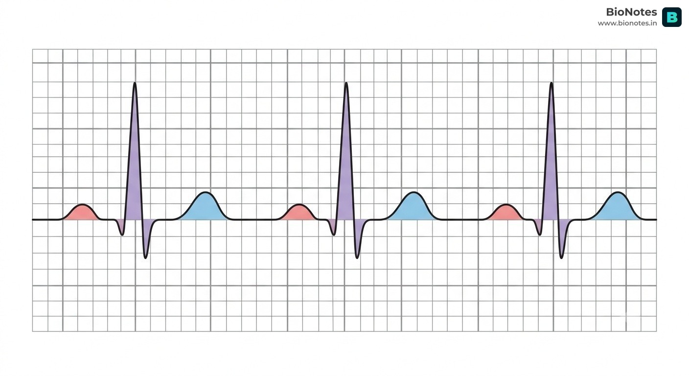
B. An ECG is a graphic record of the electrical activity of the heart during a cardiac cycle [½]. P wave — atrial depolarisation (leads to atrial contraction) [½]; QRS complex — ventricular depolarisation (leads to ventricular contraction) [½]; T wave — ventricular repolarisation (return to the normal state) [½].
(any three, 1 mark each) Body covered with feathers; forelimbs modified into wings for flight; mouth modified into a toothless beak; bones are long and hollow (pneumatic); four-chambered heart; warm-blooded (homeothermous); lay hard-shelled eggs (oviparous); lungs have air sacs.
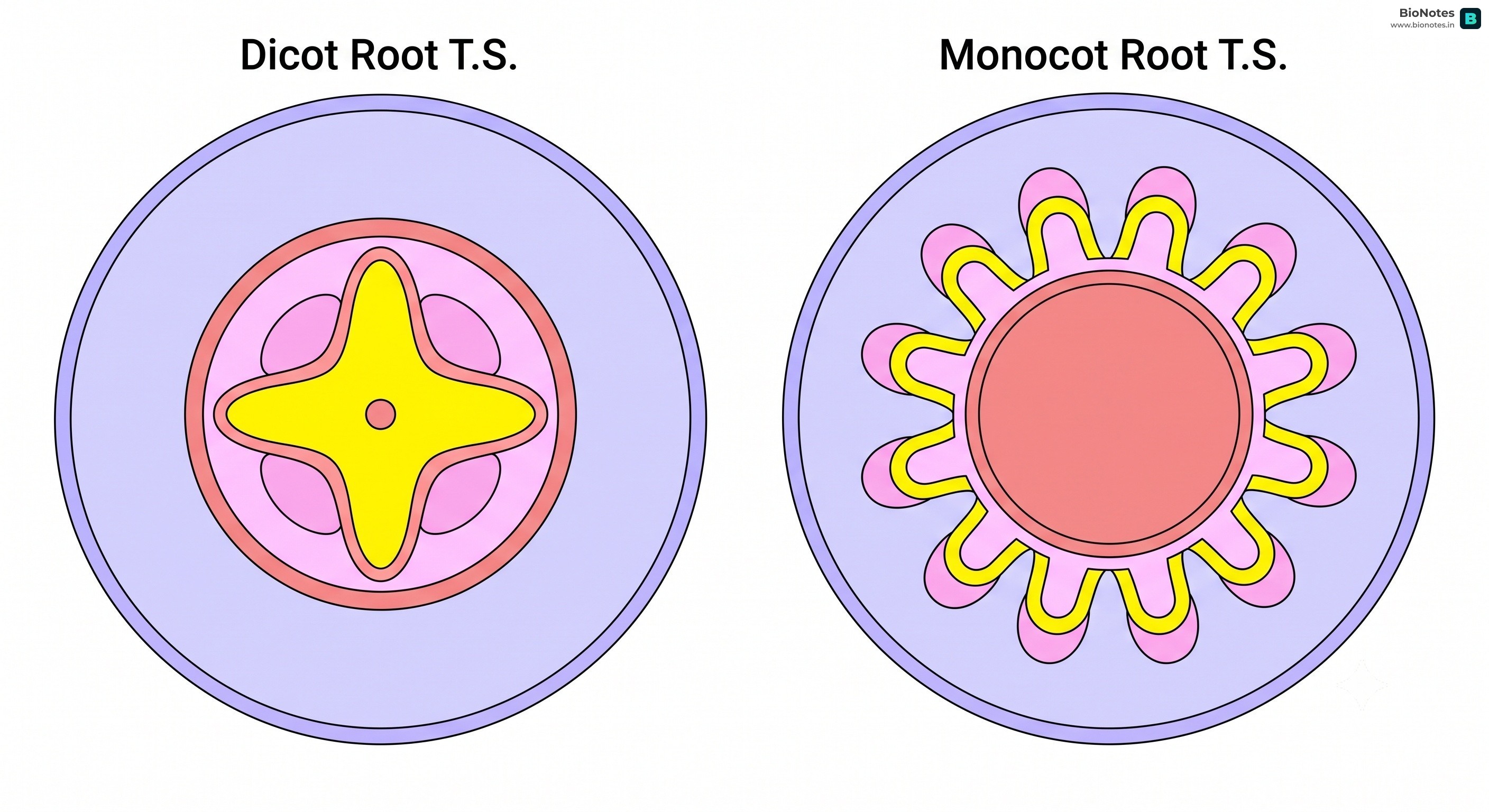
Vascular cambium develops between xylem and phloem
Vascular cambium is absent
Interactive Diagram•study Mode
||
www.bionotes.in
OR
(ii) Structure: living cells with cell walls unevenly thickened at the corners by deposits of cellulose and pectin [1]. Location: below the epidermis (hypodermis) of dicot stems and in petioles/leaf midribs [1]. Function: gives mechanical support and flexibility to growing parts [1].
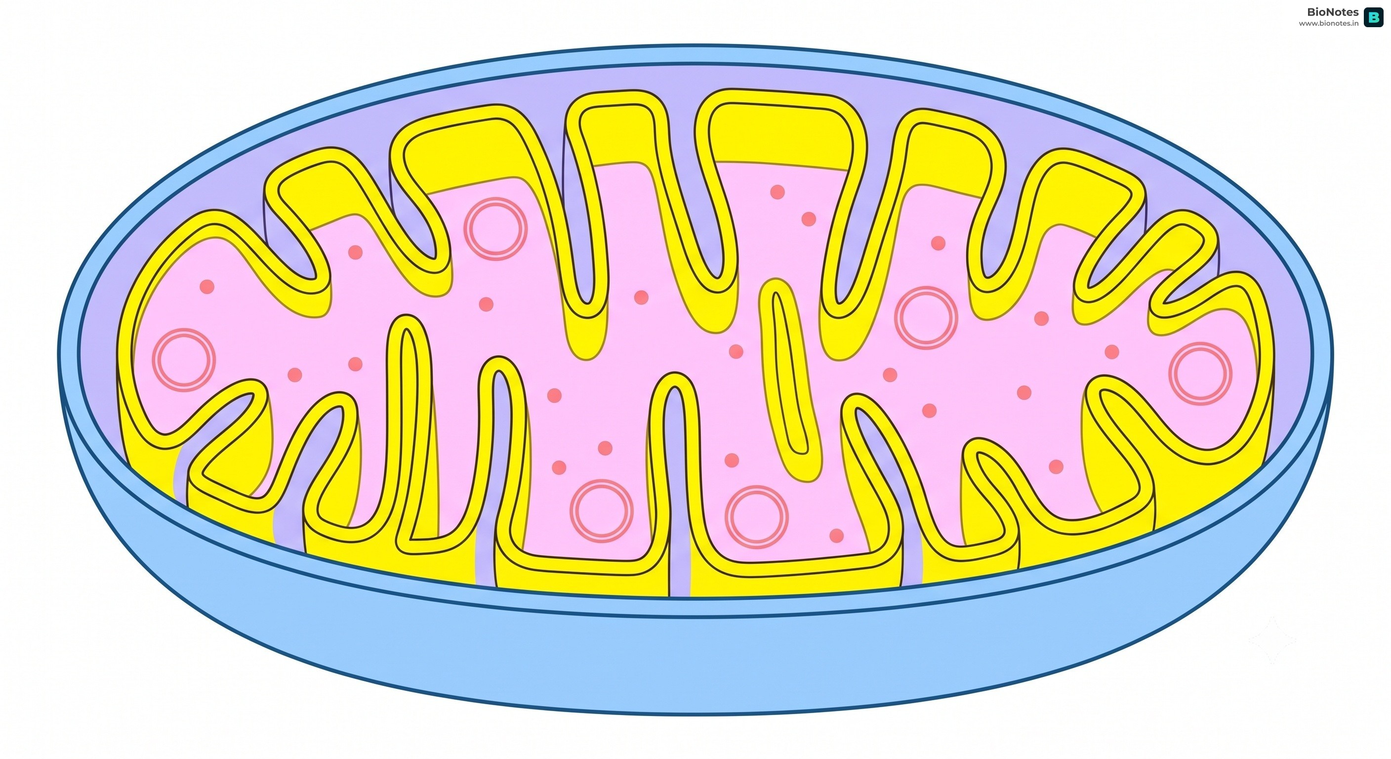
The mitochondrion is a double-membrane-bound organelle [½]. The outer membrane is smooth and permeable [½]; the inner membrane is folded into finger-like projections called cristae, which carry the ATP synthase (F₁) particles and the electron transport chain [1]; the inner space is filled with a matrix containing enzymes of the Krebs cycle, a circular DNA and ribosomes [½]. Function: the site of aerobic respiration and production of ATP — hence the "powerhouse of the cell" [½].
Photorespiration is the light-dependent uptake of oxygen and release of carbon dioxide in C₃ plants, which involves the chloroplast, peroxisome and mitochondrion [1]. When the concentration of O₂ is high and CO₂ is low, RuBisCO acts as an oxygenase and combines RuBP with O₂ instead of CO₂, forming one molecule of phosphoglycerate and one of phosphoglycolate [1]. It is wasteful because neither ATP nor NADPH is produced; instead, energy is consumed and fixed carbon is lost as CO₂, which reduces the yield of C₃ plants [1].
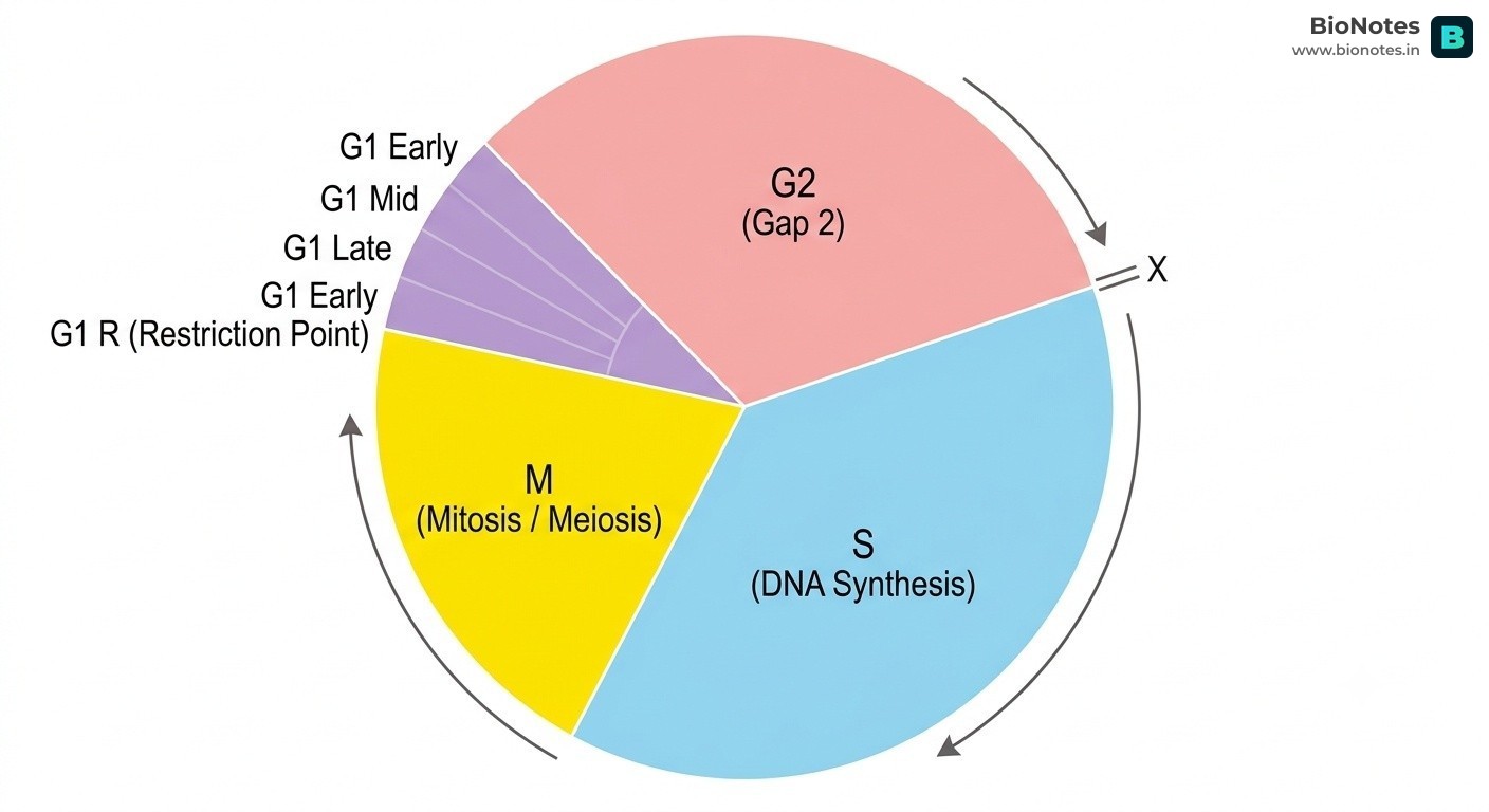
(a) [1] C-value is the amount of DNA present in the haploid set of chromosomes (haploid genome) of an organism; it is constant for a species.
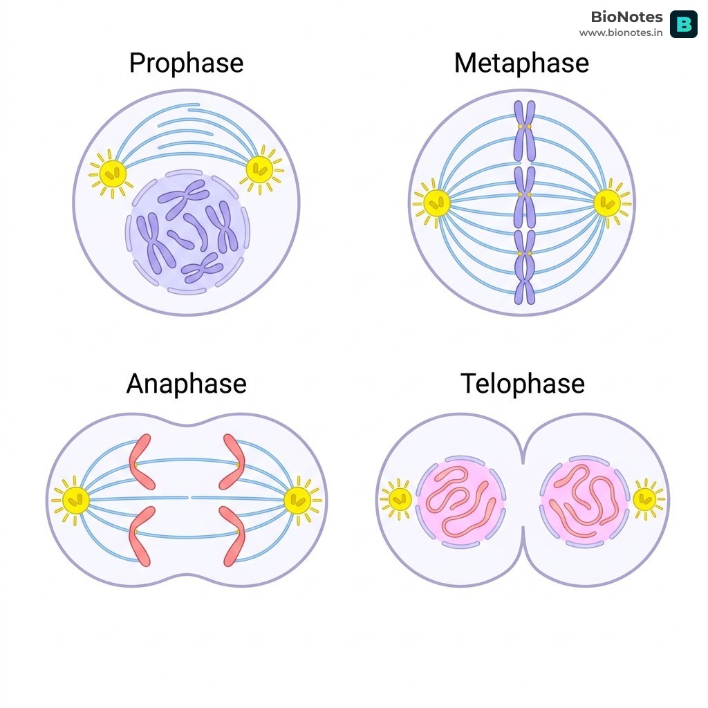
(b) [2] G₁ phase: the cell grows and is metabolically active; DNA is not replicated [½]. S phase: DNA replication takes place; the DNA content doubles but the chromosome number remains the same [1]. G₂ phase: proteins and organelles needed for division are synthesised and the cell grows further [½].
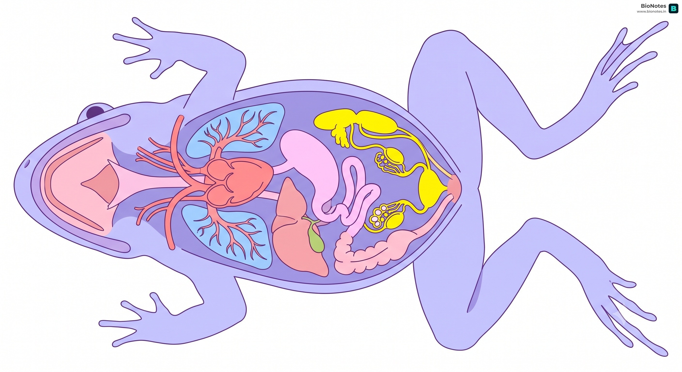
The alimentary canal of the frog is short because it is carnivorous [½]. Food passes from the mouth into the buccal cavity (which has a sticky tongue and maxillary teeth), then to the pharynx, oesophagus, stomach, small intestine (duodenum and ileum), large intestine (rectum) and finally opens into the cloaca [1½]. The digestive glands are the liver (bile, stored in the gall bladder) and the pancreas (pancreatic juice) [½]. Food is digested in the stomach by gastric juice and in the intestine by bile and pancreatic juice; absorption takes place in the intestine [½].
(a) [1] When a process is controlled by several factors, the rate of the process is limited by the factor that is nearest to its minimum value (the factor in shortest supply).
(b) [2](any two)
Cyclic photophosphorylation
Non-cyclic photophosphorylation
Involves only photosystem I
Involves both photosystem II and photosystem I
Electrons return to photosystem I
Electrons do not return; they pass to NADP⁺
Only ATP is formed
ATP and NADPH are formed
No photolysis of water; no O₂ is released
Photolysis of water occurs; O₂ is released
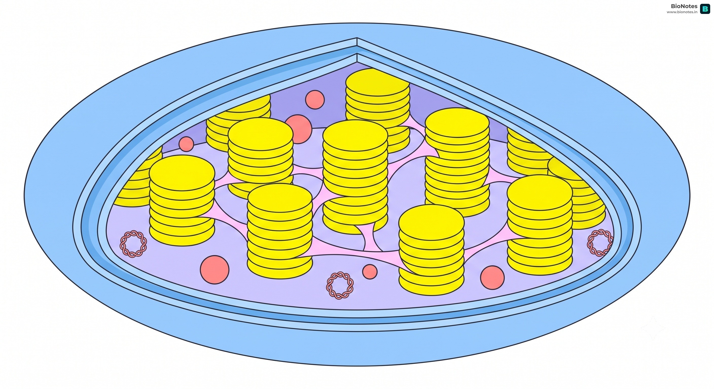
(c) [2] According to Mitchell's chemiosmotic hypothesis, ATP synthesis is linked to the development of a proton gradient across the thylakoid membrane [½]. Splitting of water inside the lumen releases H⁺ ions, and as electrons pass along the transport chain, protons are pumped from the stroma into the lumen; NADP reductase also removes H⁺ from the stroma, so the lumen becomes highly acidic [1]. The protons then flow back from the lumen to the stroma through the CF₀–CF₁ (ATP synthase) particle, and the energy released drives the synthesis of ATP from ADP and inorganic phosphate [½].
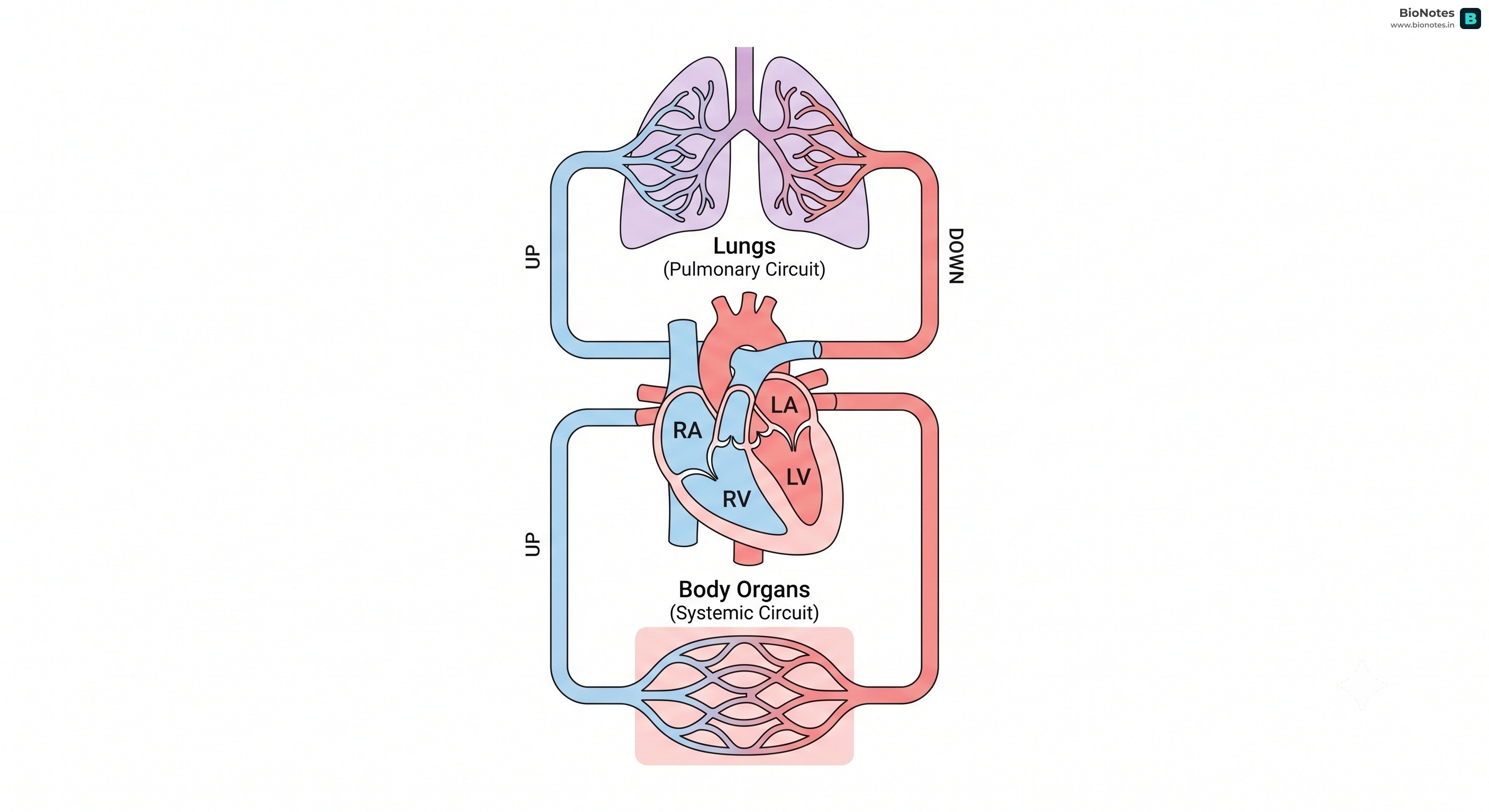
(a) [1] Stroke volume is the volume of blood pumped out by each ventricle in one heartbeat (about 70 mL).
(b) [2] Auricular systole: the two auricles contract and the blood is forced into the ventricles through the open AV valves [1]. Ventricular systole: the ventricles contract, the AV valves close (producing the 'lub' sound), the semilunar valves open and blood is pumped into the aorta (from the left ventricle) and the pulmonary artery (from the right ventricle) [1].
(c) [2] Double circulation: in one complete cycle, the blood passes twice through the heart — through the systemic circuit (left ventricle → body → right auricle) and through the pulmonary circuit (right ventricle → lungs → left auricle) [1]. Significance: oxygenated and deoxygenated blood do not mix, so tissues receive blood with a high oxygen content and the body is able to maintain a high rate of metabolism and constant body temperature [1].
Interactive Diagram•study Mode
||
www.bionotes.in
OR
(ii)
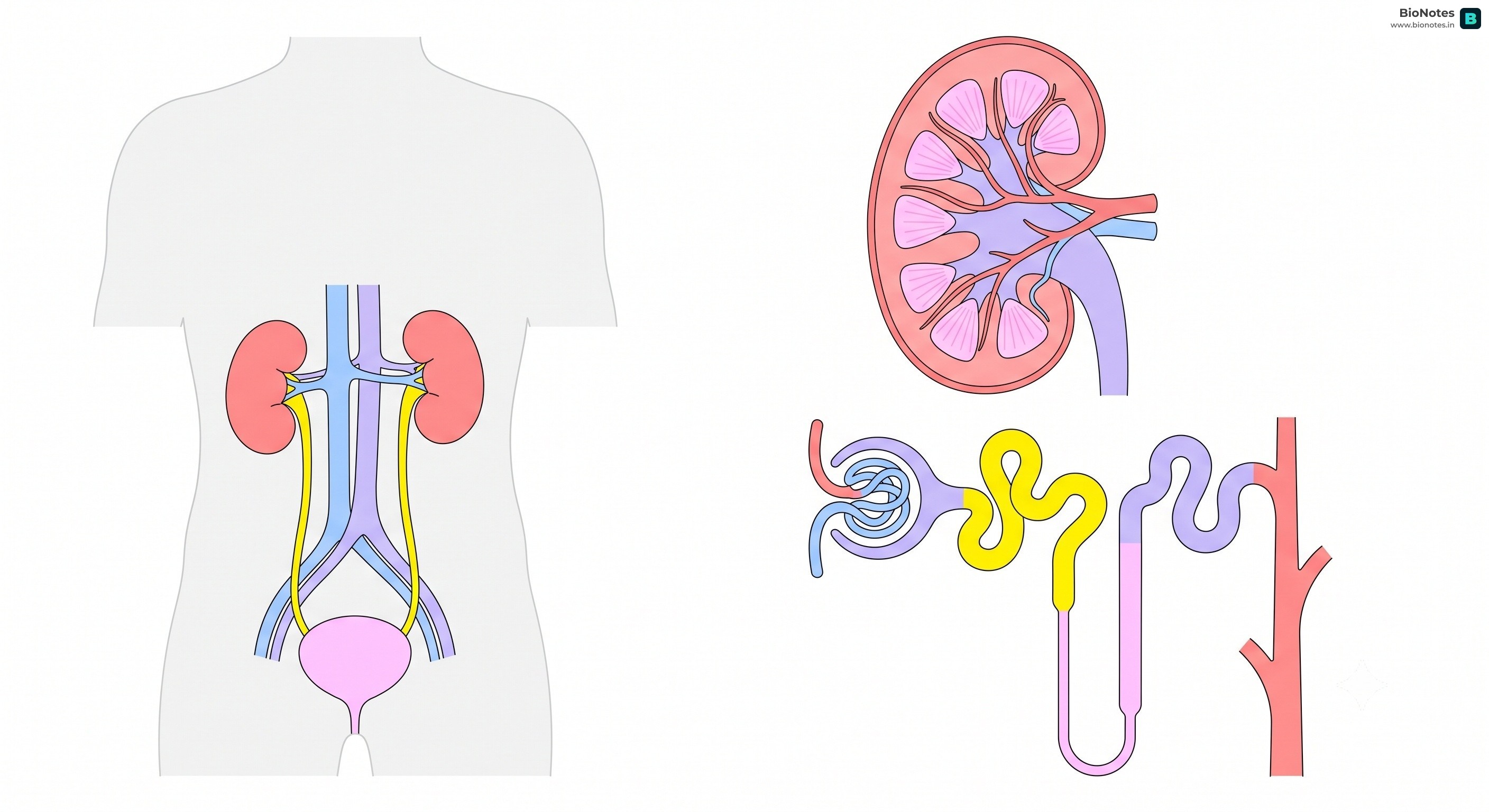
(a) [1] GFR is the volume of filtrate formed by the kidneys per minute; in a healthy adult it is about 125 mL/min (about 180 litres per day).
(b) [2] The PCT is lined by a brush-bordered cuboidal epithelium, which increases the surface area [½]. Almost all of the glucose and amino acids, about 70–80% of the electrolytes (Na⁺, K⁺, etc.) and water are reabsorbed here from the filtrate [1], partly by active and partly by passive transport [½].
(c) [2] An increase in the osmolarity of the blood is detected by osmoreceptors, which stimulate the hypothalamus to release ADH from the neurohypophysis [1]. ADH increases the permeability of the DCT and the collecting duct to water, so more water is reabsorbed and a small volume of concentrated urine is produced; lack of ADH causes diabetes insipidus [1].
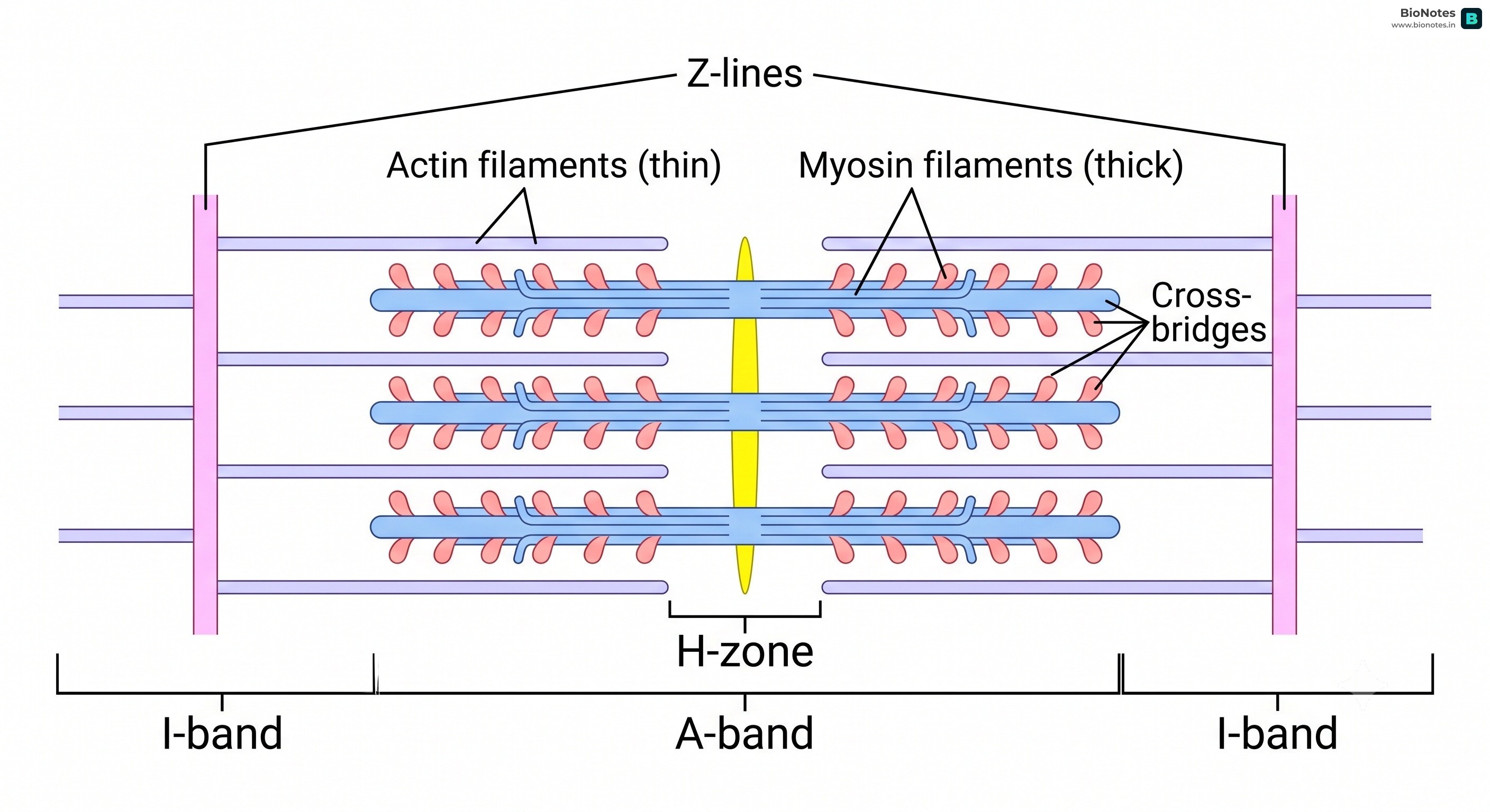
(a) [1] Actin and myosin (also acceptable: tropomyosin and troponin as regulatory proteins).
(b) [2] According to the sliding filament theory (Huxley and Niedergerke), muscle contraction takes place when the thin (actin) filaments slide over the thick (myosin) filaments [½]. On stimulation, Ca²⁺ is released from the sarcoplasmic reticulum and binds to troponin, which exposes the myosin-binding sites on actin [½]. The heads of myosin bind to actin to form cross-bridges and, using energy from ATP, pull the actin filaments towards the centre of the sarcomere [½]. As a result, the I-band and H-zone become shorter (the A-band remains the same) and the sarcomere shortens [½].