The nervous system is the master controller and communication network of the body. It is a complex, highly organized system that coordinates our actions, reflexes, and sensations, allowing us to interact with and respond to the world around us.
Watch Out! Neurobiology is highly complex. Watch out for these misconceptions:
Misconception 1:Nerves and neurons are the exact same thing.Correction: A neuron is a single nerve cell. A nerve is a bundle of thousands of neuron fibers (axons) wrapped in connective tissue, like cables in a thick wire.
Misconception 2:Reflex actions are processed by the brain.Correction: Most reflex actions (like pulling your hand away from a hot stove) are processed rapidly by the spinal cord, bypassing the brain entirely to save time.
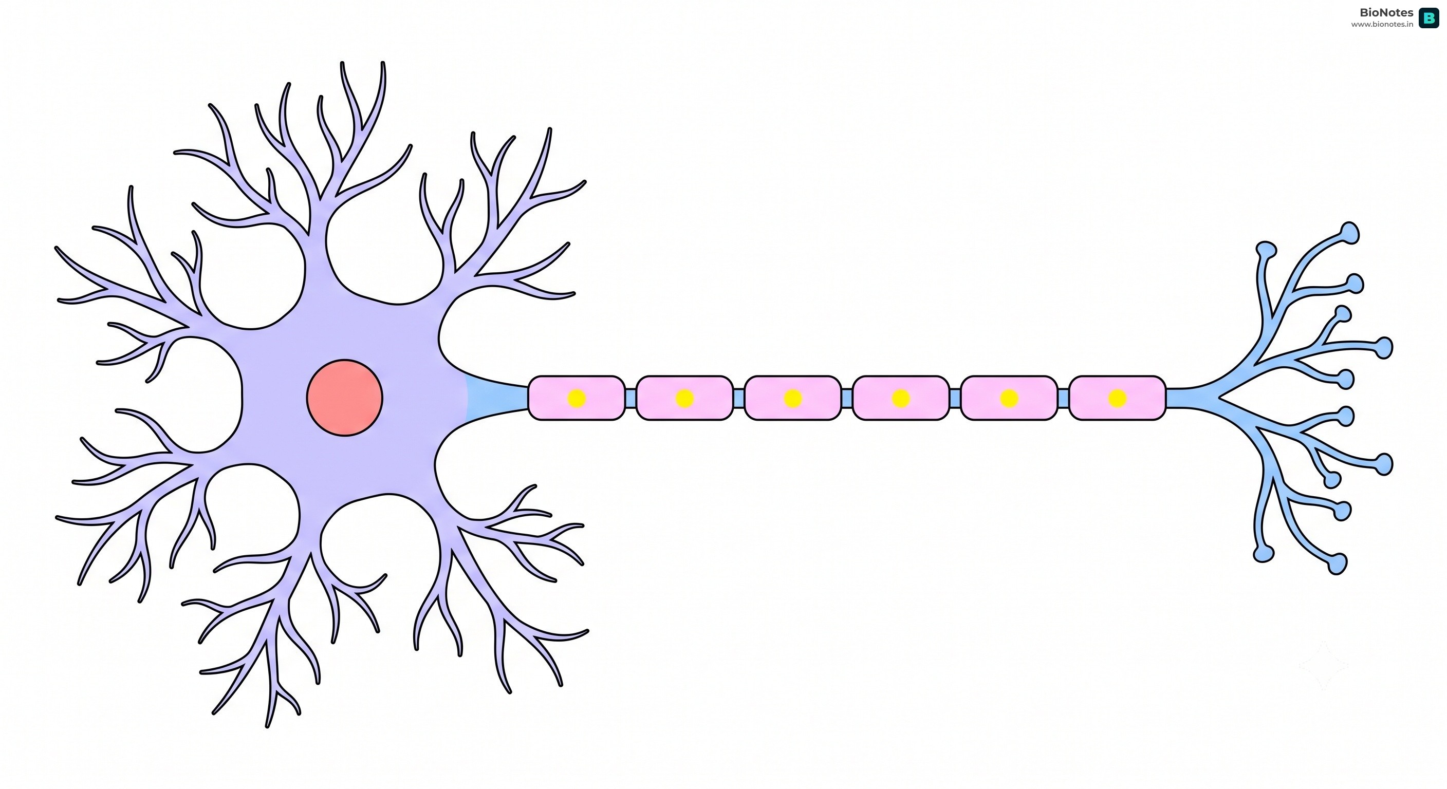
The fundamental unit of the nervous system is the neuron, a specialized cell designed to transmit information.
Cell Body (Soma or Cyton): The main part of the neuron, containing the nucleus and other organelles. It is the metabolic center of the neuron.
Dendrites: These are short, branched extensions of the cell body that receive electrochemical signals (neurotransmitters) from other neurons and transmit them towards the cell body.
Axon: A long, slender projection that carries nerve impulses away from the cell body to other neurons, muscles, or glands.
Myelin Sheath: Many axons are covered by a fatty, insulating layer called the myelin sheath, which is produced by Schwann cells. This sheath is not continuous and has gaps called Nodes of Ranvier. The myelin sheath allows for saltatory conduction, where the nerve impulse "jumps" from one node to the next, significantly increasing the speed of transmission.
Axon Terminals (Terminal Buttons): These are the branched endings of an axon. They contain synaptic vesicles filled with neurotransmitters, which are released into the synapse to transmit the signal to the next neuron.
Syllabus History & Status (Neuron & Nerve Classifications):
From ICSE 2028 onwards: Mandatory (Detailed classifications of neuron types—sensory, motor, association—and nerve types—sensory, motor, mixed—are part of the syllabus).
Up to ICSE 2027: Optional / Reference Only (General nervous system structure is studied, but these specific category distinctions are not explicitly tested).
Sensory (Afferent) Neurons: Carry impulses from the sensory receptors (sense organs) towards the central nervous system (brain and spinal cord).
Motor (Efferent) Neurons: Carry impulses from the central nervous system to the effectors (muscles or glands) to bring about a response.
Association (Interneurons or Relay Neurons): Located entirely within the central nervous system (brain/spinal cord). They connect sensory and motor neurons, processing and integrating the signals.
Nerves are bundles of individual nerve fibres (axons) enclosed in a tubular sheath.
Sensory Nerves: Contain only sensory nerve fibres. They carry sensory impulses to the CNS (e.g., Optic nerve of the eye, Auditory nerve of the ear).
Motor Nerves: Contain only motor nerve fibres. They carry response impulses from the CNS to muscles or glands (e.g., Oculomotor nerve controlling eyeball movement).
Mixed Nerves: Contain both sensory and motor nerve fibres. They can carry signals in both directions (e.g., Spinal nerves branching from the spinal cord).
The CNS is the body's command center, consisting of the brain and the spinal cord.
Competitive Edge: The Meninges (DAP)
The brain and spinal cord are protected by three tough, membranous layers called the meninges. Remember the order from outside to inside with the mnemonic DAP:
Dura mater (Outermost, tough)
Arachnoid mater (Middle, web-like)
Pia mater (Innermost, highly vascular, adheres strictly to the brain)
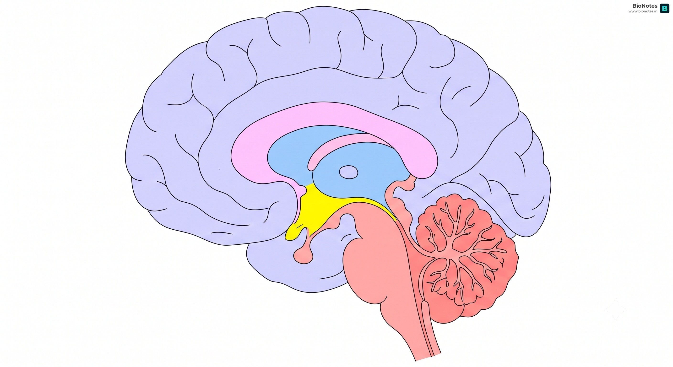
The brain is the primary control center of the nervous system, responsible for higher-order functions like thought, memory, and emotion.
Interactive Diagram•study Mode
||
www.bionotes.in
Cerebrum: The largest part of the brain, divided into two cerebral hemispheres (left and right). It is the center of intelligence, memory, consciousness, and voluntary actions.
Cerebellum: Located at the back of the brain, the cerebellum coordinates voluntary muscle movements, posture, and balance.
Medulla Oblongata: Part of the brainstem, it controls vital involuntary functions like heartbeat, breathing, and blood pressure.
Thalamus: Acts as a relay station for sensory information, directing it to the appropriate areas of the cerebrum for processing.
Hypothalamus: Located below the thalamus, it regulates body temperature, hunger, thirst, and the sleep-wake cycle. It also controls the pituitary gland, linking the nervous system to the endocrine system.
Pons: Part of the brainstem, it relays signals between the cerebrum and the cerebellum and plays a role in sleep and arousal.
Gray and White Matter: The CNS is composed of two types of tissue:
Gray Matter: Consists mainly of neuron cell bodies, dendrites, and unmyelinated axons. In the brain, it forms the outer layer (cerebral cortex), while in the spinal cord, it forms the central H-shaped core.
White Matter: Composed primarily of myelinated axons. In the brain, it is found beneath the cortex, while in the spinal cord, it surrounds the gray matter.
The spinal cord is a long, thin, tubular bundle of nervous tissue that extends from the medulla oblongata in the brainstem to the lumbar region of the vertebral column. It serves as a pathway for nerve impulses between the brain and the rest of the body and is the center for coordinating many reflexes.
The PNS consists of all the nerves that branch out from the brain and spinal cord, connecting the CNS to the rest of the body. It is further divided into the somatic and autonomic nervous systems.
Somatic Nervous System: Responsible for carrying sensory and motor information to and from the CNS. It is associated with voluntary control of body movements via skeletal muscles.
Autonomic Nervous System (ANS): Regulates involuntary bodily functions such as heart rate, digestion, respiratory rate, and pupillary response.
Syllabus History & Status (Autonomic Divisions):
From ICSE 2028 onwards: Mandatory (Detailed contrasting actions of the Sympathetic and Parasympathetic nervous systems are part of the syllabus).
Up to ICSE 2027: Optional / Reference Only (Only brief definitions of sympathetic/parasympathetic are required).
It is divided into two antagonistic (opposing) systems:
Sympathetic Nervous System: Prepares the body for emergency situations requiring quick action ("fight, flight, or fright"). It increases energy expenditure.
Parasympathetic Nervous System: Restores the body to a normal, relaxed state ("rest and digest"). It helps conserve and replenish energy.
Voluntary Actions: These are actions that are under the conscious control of the individual, such as walking, talking, and writing. They are initiated by the cerebrum.
Involuntary Actions: These are actions that are not under conscious control, such as the beating of the heart and the digestion of food. They are controlled by the autonomic nervous system.
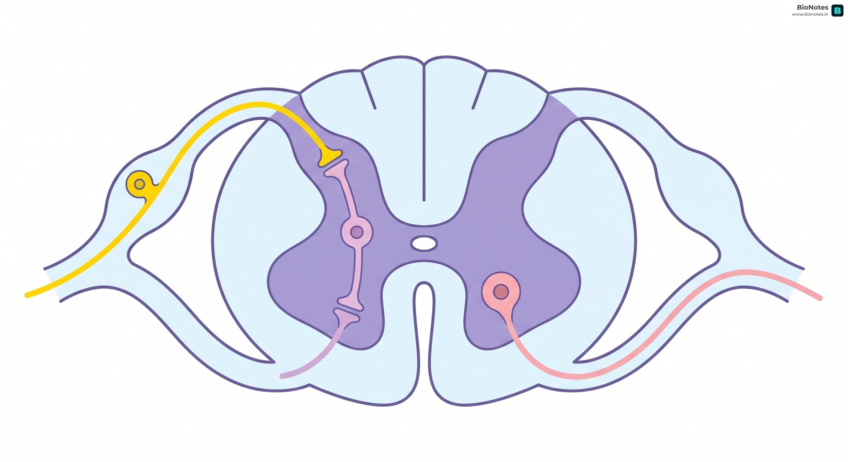
A reflex action is a rapid, involuntary, and stereotyped response to a stimulus. It is a protective mechanism that allows the body to react quickly to potentially harmful situations.
Interactive Diagram•study Mode
||
www.bionotes.in
The Bypass
Reflex actions are often processed in the spinal cord, bypassing the brain for a faster response. You pull your hand away from a hot object before your brain even registers the sensation of pain!
Reflex Arc: The neural pathway that mediates a reflex action. It typically includes:
Receptor: Detects the stimulus.
Sensory Neuron (Afferent Neuron): Transmits the nerve impulse from the receptor to the CNS.
Interneuron (Association Neuron): Located in the CNS, it processes the signal and transmits it to a motor neuron.
Motor Neuron (Efferent Neuron): Carries the nerve impulse from the CNS to the effector.
Effector: The muscle or gland that responds to the stimulus.
Natural vs. Acquired Reflexes:
Natural (Innate) Reflexes: These are reflexes that are present from birth, such as sneezing, coughing, and blinking.
Acquired (Conditioned) Reflexes: These are reflexes that are learned through experience, such as salivating at the sight of food.
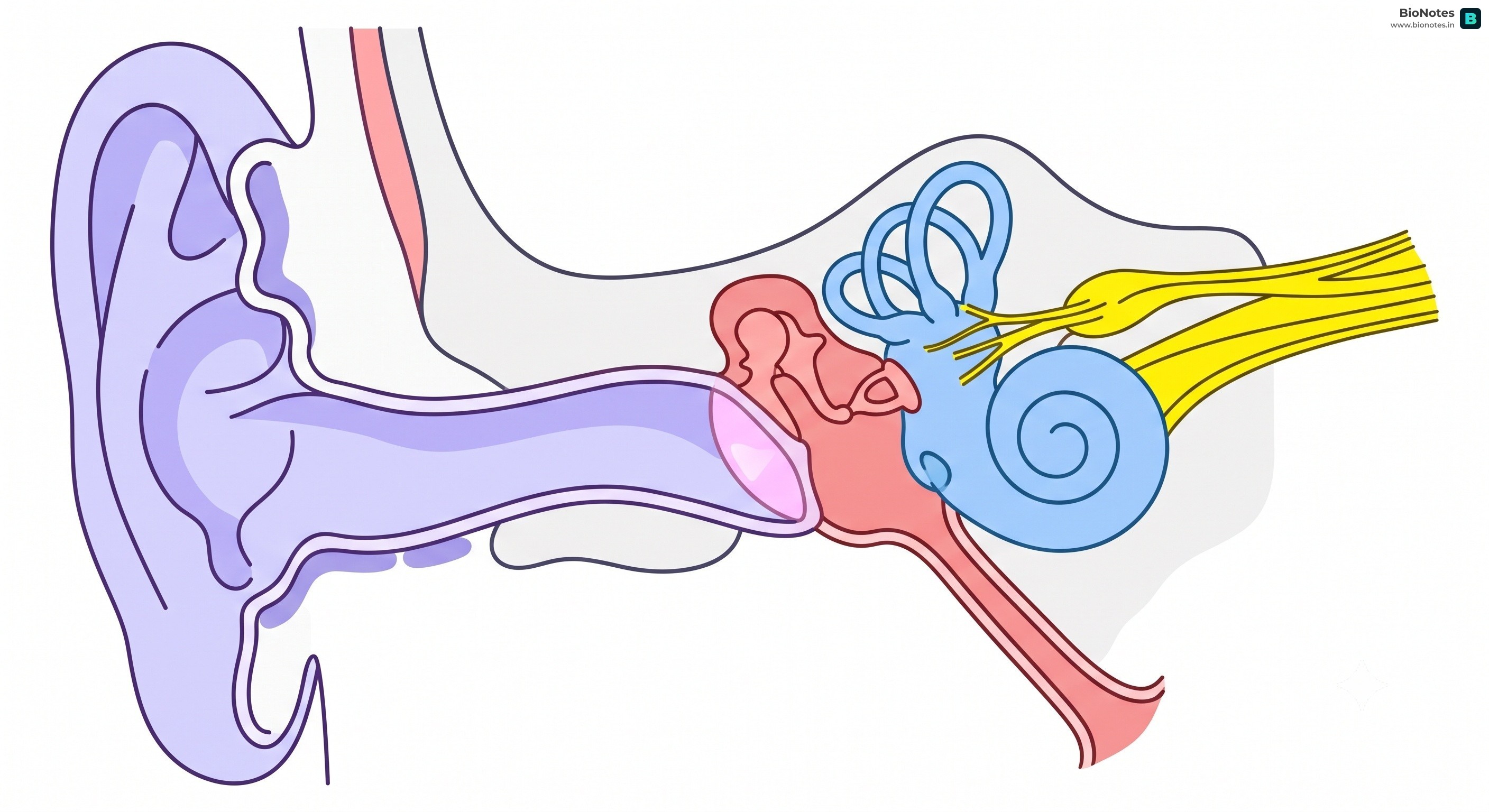
Perception of Sound: Sound waves cause the eardrum to vibrate. These vibrations are amplified by the ossicles and transmitted to the cochlea, where they are converted into nerve impulses that are sent to the brain via the auditory nerve.
Role in Balance: The vestibular system detects changes in head position and movement, sending signals to the brain to help maintain balance.
The transmission of a nerve impulse is an electrochemical process.
Syllabus History & Status (Nerve Impulse & Synapse Mechanism):
From ICSE 2028 onwards: Mandatory (Detailed electrochemical mechanism of impulse transmission and synaptic structure—synapse, synaptic transmission, and delay—are tested).
Up to ICSE 2027: Optional / Reference Only (General transmission concepts are studied, but detailed step-by-step depolarization/repolarization and synaptic structures are not explicitly required).
Resting Potential: In a resting (unstimulated) neuron, the outer side of the membrane is positively charged while the inner side is negatively charged (resting potential is about -70 mV). This polarization is maintained by the sodium-potassium pump, which actively transports 3 Na+ ions out of the cell for every 2 K+ ions it pumps in.
Action Potential (All-or-None Law): When a neuron is stimulated above a certain threshold (typically -55 mV), it fires an action potential. The strength of the response is independent of the stimulus strength, provided it exceeds the threshold (all-or-none).
Depolarization: Upon stimulation, sodium channels open, and Na+ ions rush into the cell, making the interior positive (depolarization).
Repolarization: Sodium channels close and potassium channels open, allowing K+ ions to rush out of the cell, restoring the negative charge inside (repolarization).
Hyperpolarization: The slow closing of K+ channels results in the interior temporarily becoming more negative than the resting state.
Refractory Period: The brief phase during which the neuron cannot fire another action potential, ensuring one-way propagation.
 BioNotes