The circulatory system is a complex network of organs and vessels that is responsible for transporting nutrients, oxygen, carbon dioxide, hormones, and blood cells to and from the cells in the body. This system is vital for providing nourishment, fighting diseases, stabilizing temperature and pH, and maintaining homeostasis. The circulatory system consists of two main components: the cardiovascular system (heart, blood, and blood vessels) and the lymphatic system.
Watch Out! The circulatory system is fascinating but often misunderstood:
Misconception 1:The right side of the heart pumps blue blood.Correction: Blood is never blue! Deoxygenated blood is just a darker, deeper red. Textbooks use blue and red just for illustration purposes.
Misconception 2:Arteries always carry oxygenated blood.Correction: Most do, but the pulmonary artery carries deoxygenated blood from the heart to the lungs.
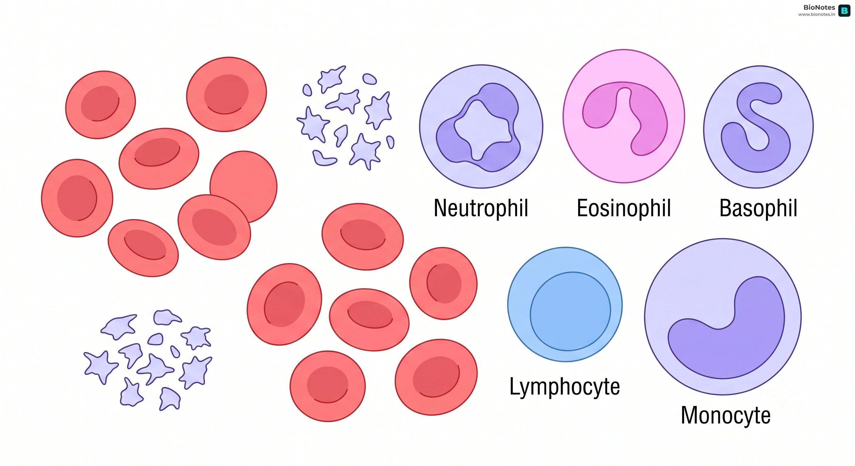
Blood is a specialized bodily fluid that delivers necessary substances to the body's cells and transports waste products away from those same cells. It is composed of plasma, red blood cells, white blood cells, and platelets.
Plasma: The liquid component of blood, constituting about 55% of its volume. It is a yellowish fluid that carries blood cells, nutrients, hormones, and waste products.
Red Blood Cells (RBCs or Erythrocytes): These are the most numerous cells in the blood. Their primary function is to transport oxygen from the lungs to the body's tissues and to carry carbon dioxide away from the tissues and back to the lungs.
Structure: RBCs are biconcave discs, which increases their surface area for oxygen exchange. In mammals, mature RBCs lack a nucleus and other organelles, which maximizes the space for hemoglobin, the protein that binds to oxygen.
Increase in Efficiency: The absence of a nucleus, mitochondria, and other organelles in mammalian RBCs allows for more space for hemoglobin, thus increasing their oxygen-carrying capacity. This also prevents the RBCs from consuming the oxygen they transport.
White Blood Cells (WBCs or Leukocytes): These cells are a crucial part of the immune system. They help the body fight off infections and other diseases. There are several types of WBCs, each with a specific function.
Platelets (Thrombocytes): These are small, irregular-shaped cell fragments that play a vital role in blood clotting. When a blood vessel is injured, platelets gather at the site and form a plug to stop the bleeding.
From ICSE 2028 onwards: Mandatory (Detailed medical terms relating to increase/decrease in blood cell counts—erythropenia, leukopenia, thrombocytopenia, polycythaemia, leucocytosis, thrombocytosis—are part of the syllabus).
Up to ICSE 2027: Optional / Reference Only (General functions of blood cells are tested, but these specific terms are not required).
Erythropenia: A condition characterized by an abnormally low count of red blood cells (erythrocytes) in the blood. It can lead to anemia and reduced oxygen transport.
Polycythaemia: A condition where there is an abnormally high count of red blood cells (erythrocytes), which increases blood viscosity (thickness) and can slow down blood flow.
Leukopenia: A condition marked by an abnormally low count of white blood cells (leukocytes), reducing the body's ability to fight off infections.
Leucocytosis: A condition characterized by a temporary, abnormally high count of white blood cells (leukocytes), typically in response to an active infection, inflammation, or physical stress.
Thrombocytopenia: A condition where there is a deficiency of blood platelets (thrombocytes), leading to impaired blood clotting and increased tendency to bleed (bruising, prolonged bleeding).
Thrombocytosis: A condition where there is an abnormally high count of platelets (thrombocytes) in the blood, which can lead to unwanted blood clot formation (thrombosis) within blood vessels.
Blood coagulation is the process by which liquid blood is converted into a semi-solid gel to form a blood clot, preventing excessive blood loss from injured vessels and sealing the wound against pathogens.
This process involves a series of cascading enzymatic reactions:
Initiation: Injured tissue cells and ruptured platelets at the site of trauma release an enzyme complex called Thromboplastin (or Thrombokinase).
Prothrombin Activation: In the presence of Calcium ions (Ca2+), Thromboplastin converts the inactive plasma protein Prothrombin into the active enzyme Thrombin.
Fibrin Polymerization: Active Thrombin acts on the soluble plasma protein Fibrinogen, converting it into insoluble, sticky, thread-like fibers called Fibrin.
Clot Formation: Fibrin threads form a dense network (mesh) at the wound site, trapping blood cells (RBCs, WBCs, and platelets) to form a physical plug or clot (scab).
Role of Vitamin K
Vitamin K is essential for the synthesis of Prothrombin in the liver. A deficiency of Vitamin K leads to delayed clotting and hemorrhaging.
Tissue Fluid (Interstitial Fluid): This is the fluid that surrounds the cells in the body. It is formed from blood plasma that leaks out of the capillaries. Tissue fluid helps to bring oxygen and nutrients to the cells and to remove waste products.
Lymph: Some of the tissue fluid returns to the capillaries, while the rest enters the lymphatic vessels, at which point it is called lymph. Lymph is a clear fluid that contains white blood cells and circulates throughout the lymphatic system, playing a crucial role in the immune response.
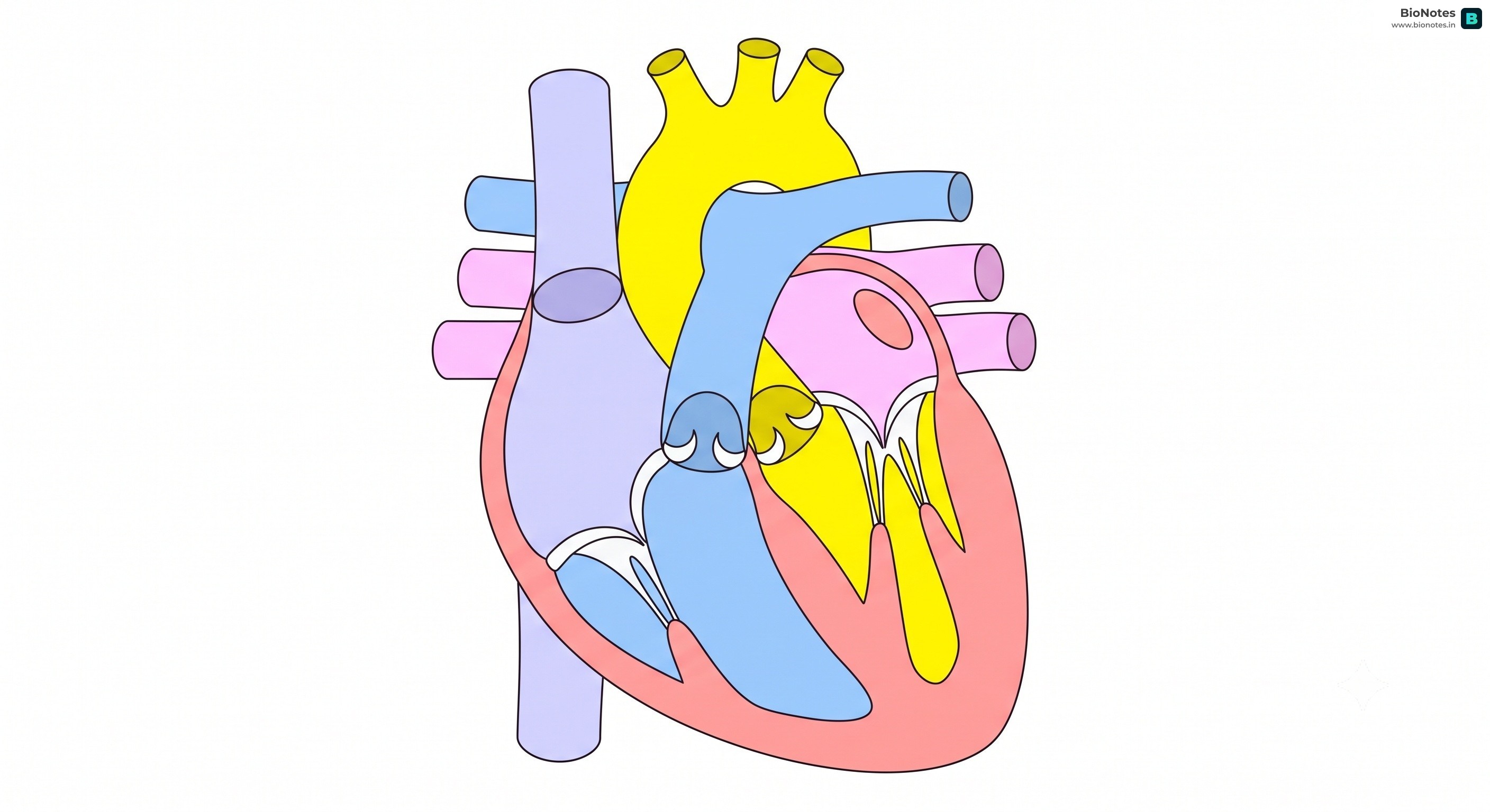
Two Atria (singular: atrium): The upper chambers that receive blood returning to the heart.
Two Ventricles: The lower chambers that pump blood out of the heart.
The right side of the heart (right atrium and right ventricle) deals with deoxygenated blood, while the left side (left atrium and left ventricle) handles oxygenated blood. Valves between the chambers and at the exit of the ventricles ensure that blood flows in only one direction.
Syllabus History & Status (SA Node - ICSE 2028 onwards):
From ICSE 2028 onwards: Mandatory (Added to the Class X syllabus: "A brief idea of natural pacemaker of heart – SA node").
Up to ICSE 2027: Optional / Reference Only (Not explicitly detailed in the syllabus).
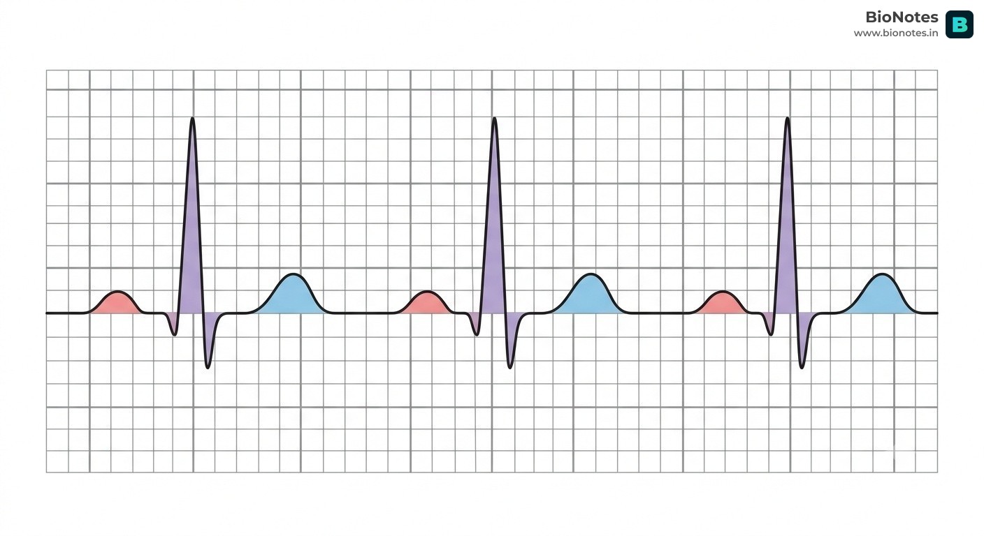
Sinoatrial Node (SA Node): The SA node is a specialized clump of cardiac muscle cells located in the upper wall of the right atrium near the opening of the superior vena cava.
Function: It is known as the natural pacemaker of the heart because it automatically generates electrical impulses (action potentials) rhythmically at a rate of 70–80 times per minute without external nervous stimulation.
Mechanism: The electrical impulse generated by the SA node spreads rapidly across the muscle walls of the atria, causing both atria to contract simultaneously (atrial systole). The impulse then travels to the Atrioventricular Node (AV node) where it pauses briefly to let the atria empty. It is then conducted down the Bundle of His and the Purkinje fibers deep into the ventricular walls, triggering a powerful ventricular contraction from the apex upwards.
The heart pumps blood in a continuous cycle known as the cardiac cycle.
Systole: The phase of the cardiac cycle when the heart muscle contracts and pumps blood from the chambers into the arteries.
Diastole: The phase of the cardiac cycle when the heart muscle relaxes and allows the chambers to fill with blood.
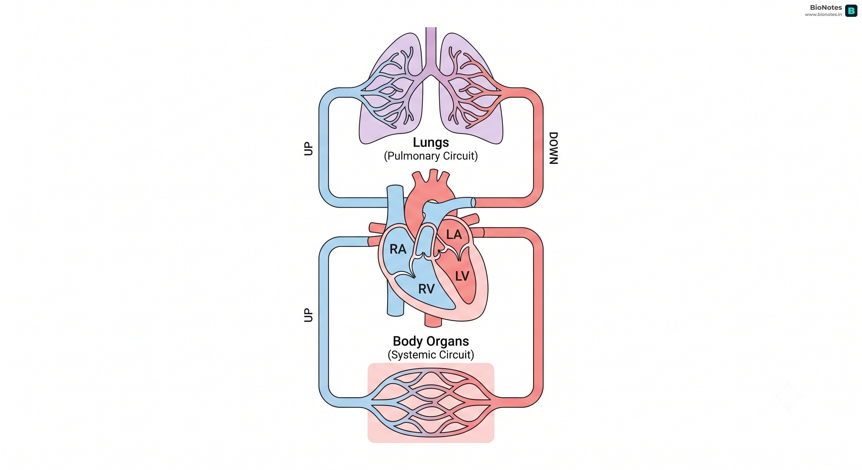
Double Circulation: In mammals, the blood passes through the heart twice for each complete circuit of the body. This is called double circulation and consists of two parts:
Interactive Diagram•study Mode
||
www.bionotes.in
Pulmonary Circulation: The right ventricle pumps deoxygenated blood to the lungs, where it gets oxygenated. The oxygenated blood then returns to the left atrium.
Systemic Circulation: The left ventricle pumps oxygenated blood to the rest of the body. The deoxygenated blood from the body then returns to the right atrium.
Vena Cava (Superior and Inferior): Bring deoxygenated blood from the body to the right atrium.
Pulmonary Veins: Bring oxygenated blood from the lungs to the left atrium.
Leaving the Heart:
Pulmonary Artery: Carries deoxygenated blood from the right ventricle to the lungs.
Aorta: Carries oxygenated blood from the left ventricle to the rest of the body.
To and from the Liver:
Hepatic Artery: Supplies oxygenated blood to the liver.
Hepatic Vein: Carries deoxygenated blood from the liver to the vena cava.
Hepatic Portal Vein: Carries nutrient-rich blood from the digestive system to the liver.
To and from the Kidneys:
Renal Artery: Supplies oxygenated blood to the kidneys.
Renal Vein: Carries deoxygenated blood from the kidneys to the vena cava.
To and from the Lungs (Pulmonary Circulation):
Syllabus History & Status (Pulmonary Vessels):
From ICSE 2028 onwards: Mandatory (Blood vessels of the lungs are explicitly added to the list of specific organ vessels to be studied).
Up to ICSE 2027: Optional / Reference Only (Only heart, liver, and kidney vessels were explicitly listed, though pulmonary vessels were studied under general circulation).
Pulmonary Artery: Carries deoxygenated blood from the right ventricle of the heart to the lungs for oxygenation.
Pulmonary Veins: Carry oxygenated blood from the lungs to the left atrium of the heart.
Pulse: The rhythmic expansion and recoil of arteries resulting from the contraction of the heart. It can be felt at various points on the body, such as the wrist and neck.
Blood Pressure (BP): The pressure exerted by the circulating blood on the walls of the blood vessels. It is usually measured in millimeters of mercury (mm Hg) and is expressed as two numbers: systolic pressure over diastolic pressure.
Heart Sounds: The "LUB" sound is caused by the closure of the atrioventricular (Tricuspid and Bicuspid) valves at the start of ventricular systole. The "DUP" sound is caused by the closure of the semilunar valves at the start of ventricular diastole.
Stroke Volume: The volume of blood pumped out by each ventricle during one heartbeat (approx. 70 mL).
Cardiac Output: The total volume of blood pumped by each ventricle per minute. Cardiac Output = Heart Rate x Stroke Volume (approx. 5 Liters/min in a healthy adult).
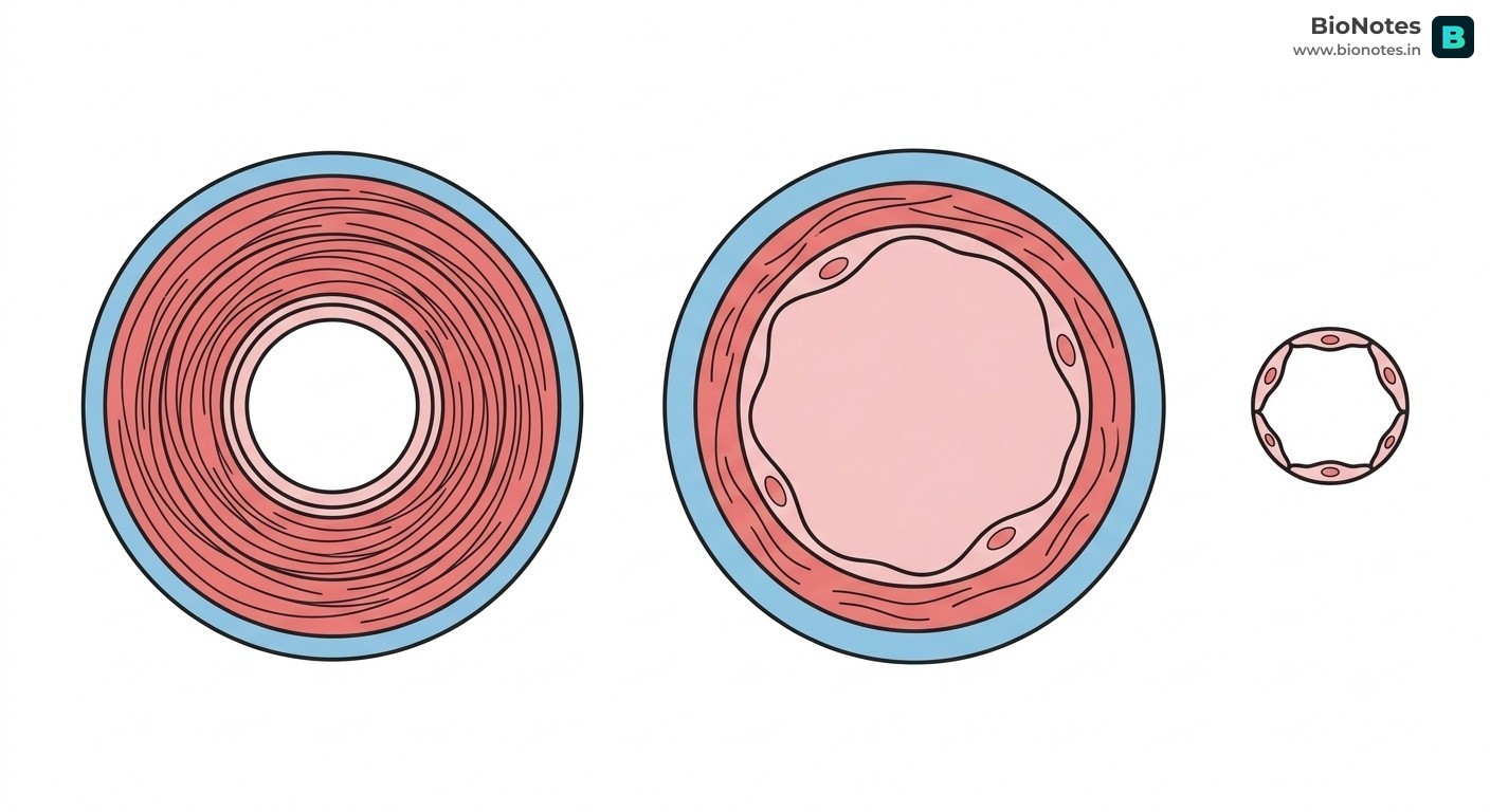
Blood vessels are the channels through which blood circulates throughout the body. There are three main types of blood vessels:
Arteries: These are thick-walled, muscular vessels that carry blood away from the heart. They are under high pressure and have elastic walls to withstand this pressure.
Veins: These vessels carry blood towards the heart. They have thinner walls than arteries and are under lower pressure. Veins have valves to prevent the backflow of blood.
Capillaries: These are the smallest blood vessels and form a network between the arteries and veins. They have very thin walls (one cell thick) to allow for the exchange of gases, nutrients, and waste products between the blood and the body's cells.
The lymphatic system is a network of tissues and organs that help rid the body of toxins, waste, and other unwanted materials. The primary function of the lymphatic system is to transport lymph, a fluid containing infection-fighting white blood cells, throughout the body.
The Rh factor is another antigen found on the surface of red blood cells. If a person has this antigen, they are Rh-positive (Rh+). If they do not, they are Rh-negative (Rh-). The Rh factor is important during pregnancy, as an Rh-negative mother carrying an Rh-positive fetus can develop antibodies that can harm the fetus.
The hepatic portal system is a unique part of the circulatory system. It is a series of veins that carry blood from the capillaries of the stomach, intestines, spleen, and pancreas to the capillaries in the liver.
Why the Liver First?
By routing nutrient-rich blood from the digestive tract to the liver before it enters general circulation, the body can regulate nutrient levels (like glucose) and detoxify harmful substances absorbed from food.
This allows the liver to process the nutrients absorbed from the digestive system before they are distributed to the rest of the body. It also allows the liver to detoxify any harmful substances that may have been absorbed.
 BioNotes